
| IMGT Web resources |
|
| Here you are: IMGT Web resources > IMGT Education > Tutorials > Immunoglobulins and B cells |
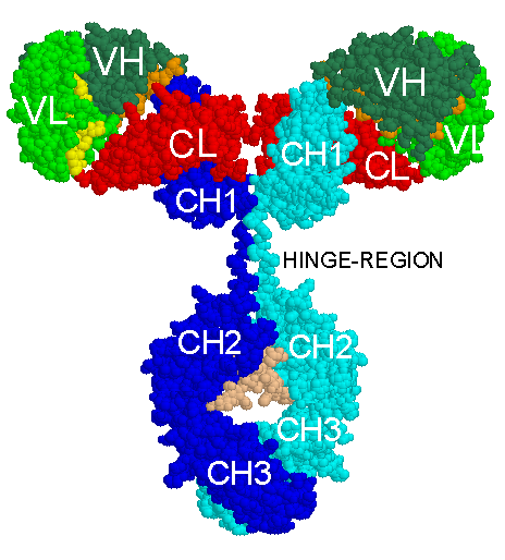
The corresponding PDB (Protein Data Bank) format file is from Mike's immunoglobulin structure function site (http://www.umass.edu/microbio/rasmol/padlan.htm).
The first chrystal 3D structure of a complete immunoglobulin is that of B12, a human IgG1, anti-HIV1, determined in 2001 (PDB code: 1hzh in IMGT/3Dstructure-DB).
This IgG1 model is described in Fig. 1 in Anatomy of the Antibody Molecule by Eduardo A. Padlan, Mol. Immunol. 31:169-217, 1994.
It is a composite model of a F(ab')2 built from the Fab from 2ig2 and an Fc fragment from 1fc2. Part of the hinge region and other details are theoretically modeled.
Created: 28/06/2001
Last updated: Friday, 06-Feb-2026 15:23:27 CET
Author: Marie-Paule Lefranc
Marie-Paule.Lefranc@igh.cnrs.fr
Editors: Elodie Foulquier, Chantal Ginestoux
IMGT Home page |
IMGT Repertoire (IG and TR) |
IMGT Repertoire (MH) |
IMGT Repertoire (RPI) |
IMGT Index |
IMGT Scientific chart |
IMGT Education |
IMGT Latest news ![]()


© Copyright 1995-2026 IMGT®, the international ImMunoGeneTics information system® | Terms of use | About us | Contact us | Citing IMGT